
MRI


Ever wondered how doctors can see inside your body without surgery? Yes, there is a medical procedure that enables medical experts to create a detailed image of your body’s organs and tissues, thereby understanding a proper treatment plan.
MRI, magnetic resonance imaging, is a medical technique used in radiology to generate images of internal body organs and tissues. This is usually a non-invasive medical procedure that uses strong radio waves and a specialized computer to create cross-sectional images of internal structures like bones and soft tissues. [1]
How an MRI Works
An MRI works by using strong magnetic fields, radio waves, and a computer to generate a detailed image of the body’s internal structures. [2]
The powerful magnetic field generated by the MRI scanner causes the atoms in your body to align in the same direction. Radio waves move the atoms out of their original position.
Using a specialized computer, radio signals are sent back, which can be analyzed by medical experts. The cross-sectional 2D images are then regenerated into a more detailed 3D image of the part of the body being diagnosed.
Instead of computed tomography (CT), MRI can be used when internal tissues are being studied. Compared to a CT scan, an MRI scan is generally considered to be better at distinguishing between different types of soft tissues and between normal and abnormal soft tissues. [3]
This medical procedure does not use ionizing radiation; hence, there is limited risk of exposure to radiation during an MRI scan.
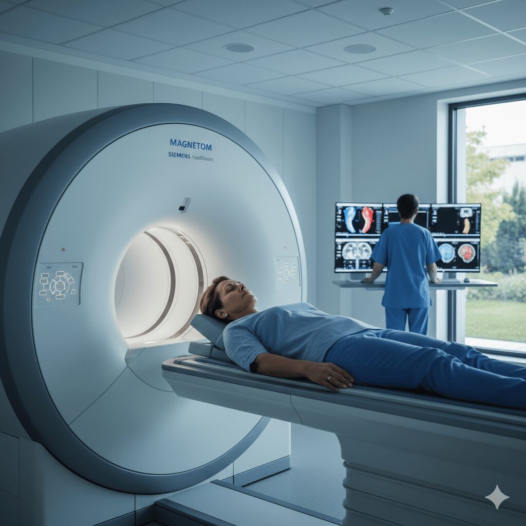
What to Expect During an MRI Scan
Before the procedure, do the following, also based on your medical professional guidelines and facility guidelines:
- Remove metallic objects. Your radiologist might ask you to remove any metallic objects from your body, be it earrings or watches. These materials can affect the quality of the scan by blurring the created image.
- Talk to your technologist about any allergies to contrast dye before the procedure.
During the scan, expect the following: [4]
- Your technologist will ask you to lie on a table to help you stay still and comfortable.
- The MRI machines produce loud and repetitive noises, which are normal during the procedure. Your healthcare provider might provide earplugs to protect your hearing.
- Expect the procedure to take about 20 minutes to over an hour. However, the duration mainly depends on the area of the body being scanned.
After the scan, your radiologist will review the images and send the report back to your healthcare professional. Your doctor will then discuss the results with you and your treatment plan.
If you received contrast dye during the procedure and notice signs of an allergic reaction like hives or shortness of breath, notify your healthcare provider immediately.
Common Uses of MRI in Medicine
MRI can be used to scan nearly every part of the head and body. This procedure is commonly used to diagnose the following conditions:
- Soft tissue injuries.
- Cardiovascular disorders.
- Brain disorders.
- Abdominal and pelvic conditions.
- Bone and joint disorders.
- Breast imaging
- Cancer detection and staging
Safety and Considerations
Your safety is usually a top priority. MRI scans are generally considered very safe as it does not expose you to ionizing radiation. Before the scan, ensure you follow every safety protocol issued by your technologists and the facility.
Always ensure all metal objects are removed from your body. An MRI scan involves strict screening for metal objects, as the powerful magnetic field can cause projectile accidents or harm you if you have certain implants.
Ensure your technologist is aware of any implanted devices such as pacemakers or insulin pumps, as many are contraindicated for MRI.
Remove any medication patches, such as glucose monitoring patches, as they can cause burns.
Bottom Line
MRI is a crucial medical procedure in modern medicine, enabling medical experts to carefully diagnose health conditions by examining the body’s internal tissues.
Do not fear the machine, as it is a powerful diagnostic tool that helps doctors understand your condition and guide them on proper treatment. Always ensure you follow your facility instructions.
Knowledge of the procedure can make the experience more comfortable and less intimidating.
Sources
- Mayo Clinic. MRI. Mayo Clinic. Published 2023. https://www.mayoclinic.org/tests-procedures/mri/about/pac-20384768
- National Institute of Biomedical imaging and Bioengineering. Magnetic Resonance Imaging (MRI). National Institute of Biomedical Imaging and Bioengineering. Published 2025. https://www.nibib.nih.gov/science-education/science-topics/magnetic-resonance-imaging-mri
- Johns Hopkins Medicine. Magnetic Resonance Imaging (MRI). Johns Hopkins Medicine. Published 2025. https://www.hopkinsmedicine.org/health/treatment-tests-and-therapies/magnetic-resonance-imaging-mri
- Stanford Medicine. Stanford Health Care. Stanfordhealthcare.org. Published 2014. https://stanfordhealthcare.org/medical-tests/m/mri/what-to-expect/during.html